
Young researchers
Access to the identity of individual cells in their original spatial context (CELL CONTEXT)
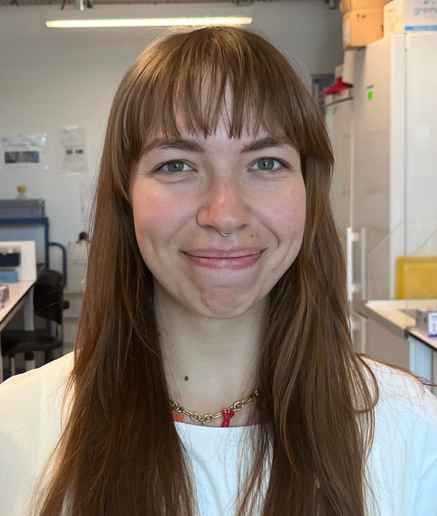
Chloé Sturmach
Photonic BioImaging (UTechS PBI), Department of Cell Biology & Infection, C2RT (Center for Technological Resources and Research)
I completed a Bachelor’s degree in Biotechnology, followed by a professional Master’s degree in Biological Engineering, where I specialized in Virology.
Read more
As part of the Cell-ID project, my role is developing automated workflows for spatial transcriptomics in thick tissue samples through the integration of fluidic systems and automated microscopy.
Because this project focuses on investigating the preservation of spatial organization of the mechanisms regulating neurogenesis in zebrafish.
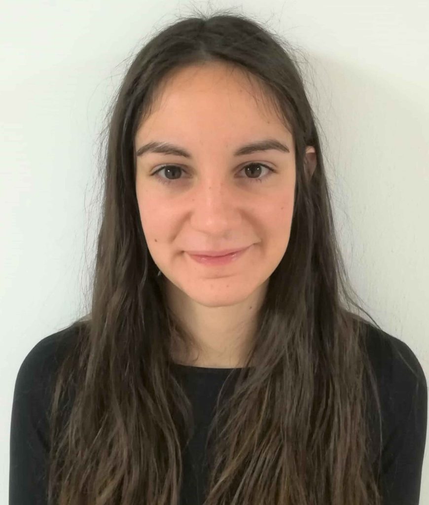
Cristina Navarrete-Hernandez
Dynamics of Genetic Information: fundamental bases and cancer (DIG-Cancer), UMR3244 (Institut Curie, CNRS, Sorbonne Université)
I completed my BSc in Biochemistry at the Universidad Autónoma de Madrid (UAM, Madrid, Spain), followed by an MSc in Genetics and Genomics at the Universitat de Barcelona (UB, Barcelona, Spain). I then spent two years as a Research Assistant at the International Institute of Molecular and Cell Biology (IIMCB, Warsaw, Poland), before returning to Barcelona to pursue my PhD on chromatin evolution at the Centre for Genomic Regulation (CRG, Barcelona, Spain). I am currently a postdoctoral researcher in Céline Vallot’s lab at Institut Curie (Paris).
Read more
I joined Céline Vallot’s lab at Institut Curie in October 2025, working on the chromatin states that define cancer cell identity through multimodal single-cell epigenomics. My project aims to decode how combinations of epigenetic marks distinguish normal from cancer cells, and how these landscapes evolve during early tumor initiation in breast cancer.
Since the early stages of my scientific career, I have been driven by a strong interest in epigenomics and omics method development, which shaped my PhD, where I developed genomic methods to study chromatin evolution across eukaryotes. As a postdoctoral fellow, I aim to extend this interest to a translational context, developing single-cell multimodal approaches to decode chromatin landscapes in cancer and understand the epigenetic processes underlying tumor initiation.
Specialized experimental systems (CELL EXP)
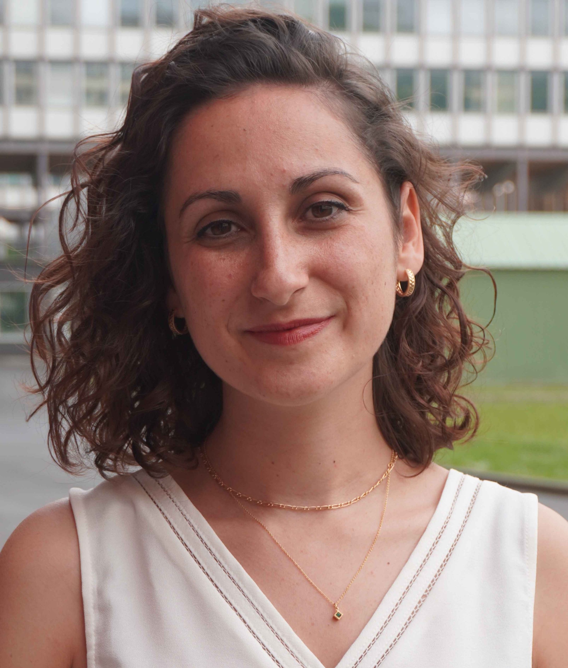
Ludovica Brunetti
Cell Biology and Cancer (UMR144) Cell Biology of Mammalian Neurogenesis (Institut Curie)
After completing a Master’s degree in Cellular and Molecular Biology at the University of Rome, I moved to Paris to pursue a PhD in Neurodevelopmental Biology at Sorbonne University. After my PhD, in 2025 I joined Alexandre Baffet’s lab as a postdoctoral researcher within the PEPR Cell-ID research program, where I now study how developmental cell lineages are established in the human cerebellum and how their disruption can lead to pediatric cerebellar tumors.
Read more
My postdoctoral project focuses on understanding how developmental cell trajectories are established in the human cerebellum and how their disruption can lead to medulloblastoma, the most common malignant brain tumor in children. To address this question, I use complementary experimental models, including human cerebellar organoids and human fetal tissue. By combining these models with live/fixed correlative microscopy, genetic manipulation for oncogene overexpression, and single-cell multi-omics approaches, I aim to reconstruct lineage trajectories during development and investigate how they respond to genetic perturbations. This work will help uncover the earliest cellular and molecular events that drive lineage derailment and tumor initiation, providing new insights into the developmental origins of aggressive medulloblastoma subgroups.
The human cerebellum is a fascinating and understudied brain structure that represents only 10% of total brain mass but contains nearly 80% of all neurons in the human brain. Primary progenitor zones are uniquely expanded and prolonged in the human cerebellum compared to other mammals. While these developmental features may have contributed to the evolutionary expansion of our cognitive abilities, they also create vulnerabilities that may predispose cells to malignant transformation.
Medulloblastoma, the most common malignant brain tumor in children, is thought to arise from specific progenitor populations of the developing cerebellum. However, we still do not know precisely when these cells diverge from their normal developmental trajectory or which molecular events drive their progression toward cancer. Understanding these early steps is essential for identifying the origins of the disease and may ultimately help improve early detection, risk stratification, and therapeutic strategies for patients.
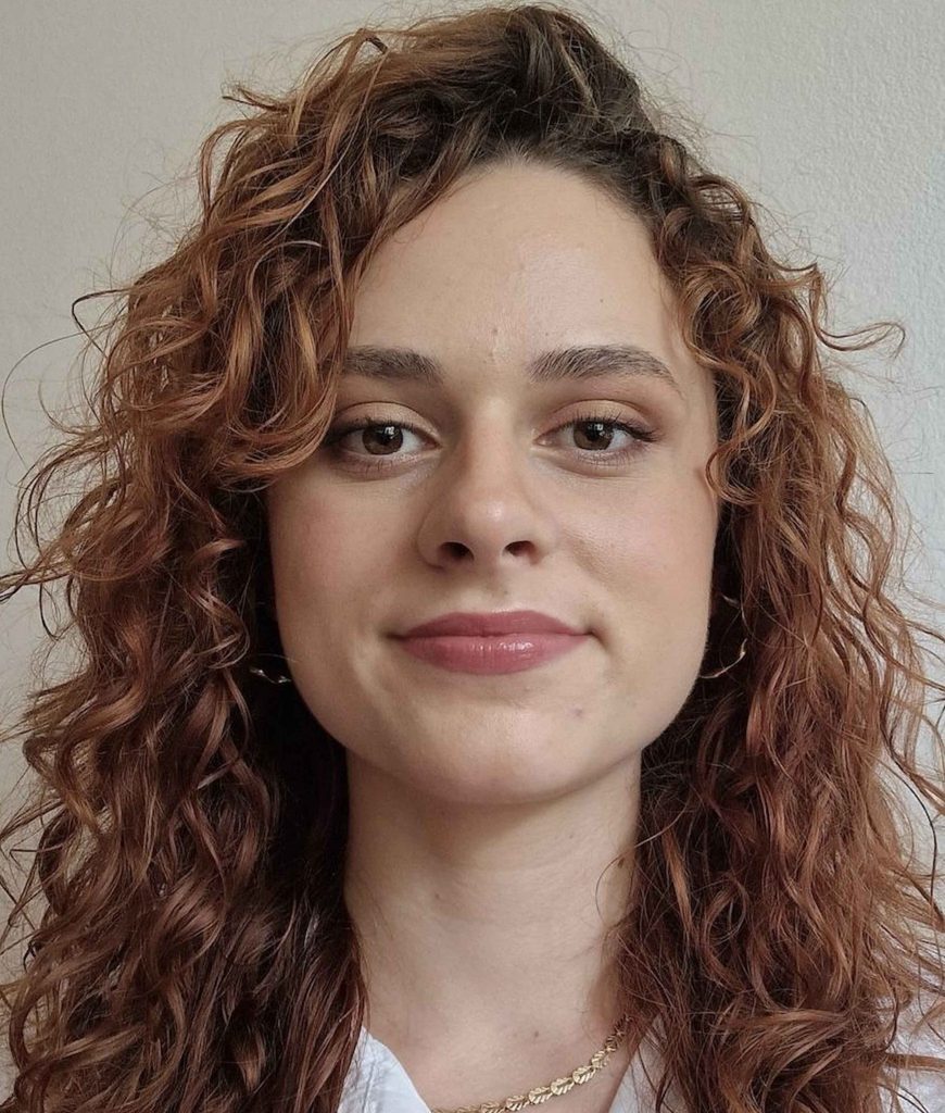
Ana Boranijasevic
uclear Dynamics (CNRS UMR UMR3664), Chromatin Dynamics Team (Institut Curie, Institut Pasteur, CNRS)
My academic journey began at the University of Belgrade, where I studied Molecular Biology and Physiology, before moving to Paris to earn my Master’s degree in Neuroscience from Université Paris Cité. Building on that foundation, I am now completing a dual-mentored PhD at two of France’s premier research institutes: Institut Curie and Institut Pasteur.
Read more
For my doctoral research, I am investigating the epigenetic mechanisms underlying pediatric brain tumors, with a specific focus on the role of histone H3 variants and their dedicated histone chaperones. Because pediatric brain cancers are often driven by profound epigenetic dysregulation rather than high mutational burdens, understanding how these chromatin components dictate cellular identity and gene expression is critical.
My project bridges the gap between experimental biology and computational analysis. On the wet-lab side, I am exploring chromatin dynamics in normal and derailed human brain development, exploiting organoids as a model system. By the bioinformatic integration of multi-omic datasets in the dry-lab, I aim to map altered epigenetic landscapes driven by H3 variant abnormalities.
Because it perfectly merges my deep fascination with neuroscience and chromatin organization. The opportunity to unravel the epigenetic complexities of the developing brain, specifically the role of histone H3, knowing this work could eventually translate into treatments for children facing devastating tumors, is incredibly motivating.

Carla Rodriguez Villa
Coming soon
Portrait Coming Soon
Read more
…

Asna Abdou
Coming soon
Portrait Coming Soon
Read more
…
Data processing for disease interception (DATA MED)
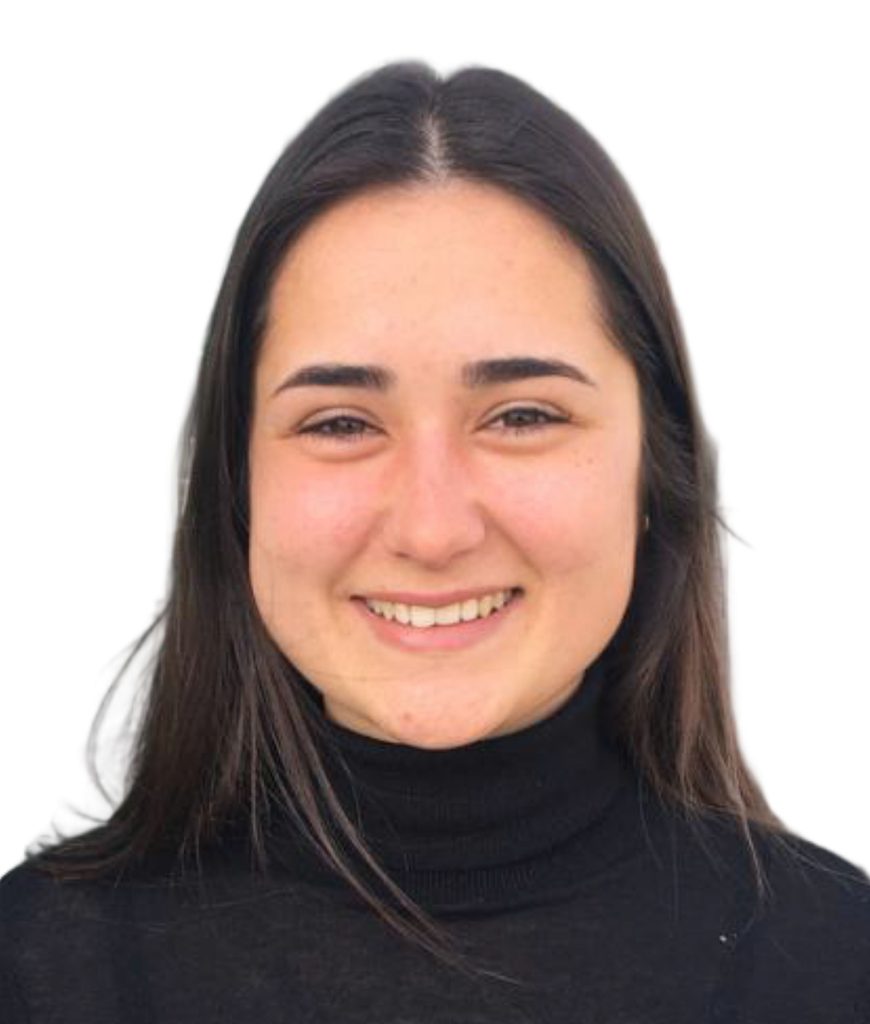
Naomie Pont
Zebrafish neurogenetics Unit / Stem cells and development Unit (Institut Pasteur, CNRS)
I completed both my bachelor’s in Life Sciences Engineering and my master’s in Computational Biology at the École Polytechnique Fédérale de Lausanne (EPFL). For my master’s thesis, I joined a Spanish startup where I contributed to developing machine learning-based pipelines for cancer detection in liquid biopsy. I then returned to France to take on a role as a Bioinformatics Research Engineer at the Institut Pasteur in Paris.
Read more
As part of the Cell-ID project, my role is developing and benchmarking analysis pipelines for spatial transcriptomics data, comparing technologies such as Stereoseq, Vizgen, and Visium HD. Especially, I am responsible for comparing the performance of the different methods and identifying the key criteria that guide the choice of one method over another.
Since the start of my contract, I have developed an analysis pipeline for Visium HD human embryo head sections to derive signatures of extraocular muscles. I am currently working on Xenium zebrafish brain sections to investigate how spatial organization of neural stem cells correlates with their quiescence depth.
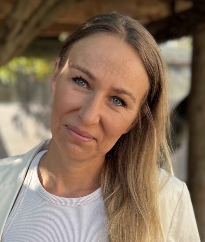
Agata Banach-Latapy
Developmental and Stem Cell Biology (UMR3738), Structures and signals in the neurogenic niche (Institut Pasteur, CNRS)
I obtained my first master’s degree in biotechnology from a University in Krakow. After completing my second master in cell biology in Paris I did a PhD in cancer cell biology. During my post-docs I specialised in neurodevelopment.
Read more
My postdoctoral project aims to identify the microenvironmental factors facilitating Atypical Teratoid/Rhabdoid Tumour (AT/RT) initiation and progression in infants. To better understand the extrinsic role of the cellular microenvironment in tumour transition and growth rate regulation, I use Drosophila Melanogaster as a model.
Using complex genetic manipulations, advanced imaging and omics approaches I want to uncover molecular mechanisms underlaying cancer cell interactions with host cell populations, hoping to identify early markers and events of the tumorigenic process.
Rhabdoid tumours are aggressive fast-growing tumours with poor prognosis. As theses tumours are rare, limited data are available to fully understand its origin and specificities to adapt therapeutical approaches for better survival rate. This study will enable better understanding of the fundamental mechanisms involved at early stages of tumour development and underlying their aggressiveness, a knowledge essential for better management of these paediatric brain tumours.
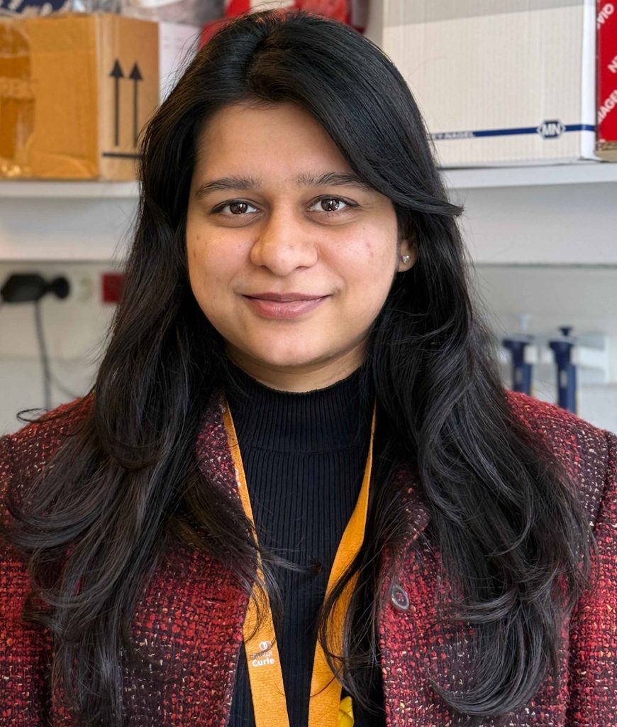
Shireen Shajahan
Childrens’ ONCology rEseaRch uniT (CONCERT), (Institut Curie, Inserm)
After a bachelor’s degree in Biochemistry (University of Delhi, India) and a master’s in Biotechnology (JMI, Delhi, India), I moved to Paris to complete a PhD in Cellular and Developmental Biology at Institut Pasteur/Sorbonne Université. Currently, I am working as a postdoctoral researcher in Franck Bourdeaut’s team within the PEPR Cell-ID program.
Read more
My postdoctoral project focuses on identifying the cell of origin of Atypical teratoid/rhabdoid tumours (AT/RT), an aggressive rare tumor of the brain in children, and to study epigenetic alterations that lead this cell type to derail from normal development and advance towards a cancerous fate. To address this question, I use approaches like single-nuclei RNA seq on patient samples to study tumor’s differentiation trajectory as compared to the normal brain. In addition, I take advantage of ATRT mouse model developed by my team that recapitulates various features of human tumors. Using techniques like sequencing-based spatial transcriptomics, I aim at identifying the earliest tumor initiating cell and following its differentiation during mouse development and tumor evolution. This work will yield important insights into the development of these tumours in young infants and provide the foundational knowledge needed to devise novel and specific treatment strategies.
ATRT are aggressive embryonal tumor found in young infants and exhibit a poor prognosis. Although it is driven by mutation is just a single gene called SMARCB1,this tumor is much more complex in its biology. It has various subtypes which differ markedly in their molecular profile (transcriptome, epigenome) and clinical features. This highlights that the unique characteristics of different ATRT subtypes are likely rooted in the cell of origin of these tumors rather than the driving mutation. Understanding how normal development is disrupted leading to formation of these tumors is crucial for designing novel and targeted therapy, patient stratification and better diagnostics.
Maya Arnould
Coming soon
Portrait Coming Soon
Read more
…
Vincent Noel
Coming soon
Portrait Coming Soon
Read more
…
Saran Pankaew
Coming soon
Portrait Coming Soon
Read more
…
Yassin Ajlil
Coming soon
Portrait Coming Soon
Read more
…
Ambre Petitalot
Coming soon
Portrait Coming Soon
Read more
…